News Story
3D Cell Culture Models for Drug PK, Safety, Efficacy Assessment
The U.S. Food and Drug Administration (FDA) and the University of Maryland Center for Excellence in Regulatory Science and Innovation (M-CERSI) co-hosted the virtual “3D Cell Culture Models for Drug PK, Safety, and Efficacy Assessment” workshop on August 14, 2020. This meeting drew 360 attendees, including stakeholders from the FDA, academia, and industry, as well as participants around the world.
This four-hour meeting aimed to address an unmet need in developing better models to identify and accurately predict drug risks and efficacy. It was designed to advance regulatory science by modernizing toxicology to enhance product safety, promote the use of novel cell and tissue models that better represent human drug responses, promote a better understanding of toxicity mechanisms by evaluating physiologically relevant 3D models at multiple molecular biological levels, and improve the ability of non-clinical models/tests for early risk assessment.
To ensure the safety and efficacy of drugs, the FDA published a number of guidances in the past decade providing recommendations to pharmaceutical industry for assessing drug-drug interactions, drug-induced liver toxicity, as well as investigational new drug (IND)'s safety in general. Collectively, preclinical testing has served as a foundation for evaluation of the potential risks and effectiveness of IND drug products in humans. Nevertheless, current two dimensional-based in vitro cell culture systems cannot accurately depict and simulate the rich environment and complex processes observed in vivo, while animal studies present significant drawbacks such as inherited species-specific differences and low throughout scales for increased demands. To improve the preclinical prediction of drug safety and efficacy, researchers continue to develop better models to evaluate and promote the use of improved cell and tissue based assays for more accurate representation of human susceptibility to drug response.
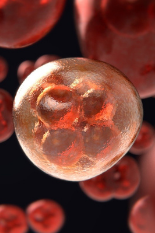
The emerging three-dimensional (3D) cell culture models appear to be a more accurate representation of the natural environment experienced by the cells under physiological and/or pathophysiological conditions and offer great potentials in assessing drug disposition and pharmacokinetics that influence drug safety and efficacy at an early stage of drug development. Currently, there are many different types of 3D culture systems, from simple spheroids to more complicated organs-on-chips and from single-cell type static 3D models to cell-coculture 3D models equipped with microfluidic flow control, each kind of them provides different advantages and disadvantages. It is noteworthy that 3D culture model is a relatively new technique that researchers have not yet fully grasped their phenotypical performance and potential implications. How well these different 3D models have advanced and which of them are suitable models that best reflecting physiologically relevant human situations are critical for improving the quality of drug safety and efficacy assessment. This educational workshop focuses on an area of regulatory science identified by FDA and University of Maryland scientists in developing better models to identify and accurately predict drug risks and efficacy.
The August 14 workshop was split into four sessions, featuring a series of ten-minute presentations in addition to two fifty-minute panel discussions.
Session One - “3D Models in Drug Safety and Risk Assessment” - featured three deliveries:
-
Overview of 3D Cellular Model Research at FDA (Suzy Fitzpatrick, Ph.D.)
-
Organ-on-Chips Application in Drug PK and Efficacy Evaluation (Lorna Ewart, Ph.D.)
-
Considerations About 3D Culture Models for Nonclinical Safety Evaluation (Ronald Wange, Ph.D., and Paul Brown, Ph.D.)
Session Two - “3D in vitro Liver Models for DILI” - included three presentations:
-
Liver-on-Chip Model for Toxicity and PK (Alexandre Ribeiro, Ph.D.)
-
HepaRG 3D Spheroids in Comparison to 2D Models (Steve Ferguson, Ph.D.)
-
Predicting DILI Risk Using Hepatic Spheroid Co-Culture Models (Will Proctor, Ph.D.)
The second session concluded with a panelist discussion between all of the speakers and moderators from the first two sessions, as well as Shannon Mumenthaler.
Session Three - Multi-Organ 3D Models Intestine, Liver, and Beyond - featured three deliveries:
-
Novel In Vitro Hepatic and Enteric Technologies for Drug Metabolism, DDI, and Safety Evaluation (Albert Li, Ph.D., MBA)
-
Intestinal Oganoids: An Excellent Ex Vivo Model for Mucosal Regeneration and Defense (Jian-Ying Wang, M.D., Ph.D.)
-
Use of Vascularized Human Kidney Proximal Tubule Microphysiological System and PBPK Modeling to Predict Renal Clearance in Subjects with Variable Kidney Function (Nina Isoherranen, Ph.D.)
Session Four - “3D Spheroids/Organoids for Disease Modeling” - included two presentations:
-
Kidney Organoids for Disease Modeling and HTS (Benjamin Freedman, Ph.D.)
-
Neuronal Multi-organ-on-Chip Models for Disease Modeling and Risk Assessment (James Hickman, Ph.D.)
The meeting concluded with another panelist discussion, featuring all of the speakers and moderators from sessions three and four and Scott Heyward.
This workshop is supported by the Food and Drug Administration (FDA) of the U.S. Department of Health and Human Services (HHS) as part of a financial assistance award U01FD005946 totaling $5,000 with 100 percent funded by FDA/HHS. The contents are those of the author(s) and do not necessarily represent the official views of, nor an endorsement, by FDA/HHS, or the U.S. Government.
Published September 21, 2020
Related Stories
Stories / October 29, 2020
M-CERSI Accepting Applications for Workshops, Research...

Stories / October 29, 2020
M-CERSI Accepting Regulatory Science Innovation Submissions
Stories / November 29, 2016
"America's Got Regulatory Science Talent" Competition Call for...

Stories / December 21, 2022
M-CERSI Now Accepting 2023 Regulatory Science Competition...

Stories / February 17, 2022
M-CERSI Celebrates 10th Annual Regulatory Science Competition
Stories / December 3, 2021
Polli Recognized for Regulatory Excellence in Education

Stories / November 12, 2021
FDA, CERSI Accepting Applications for 2022 Digital Health...
Stories / November 1, 2021
M-CERSI Accepting Regulatory Science Competition Applications
Stories / October 25, 2021
Improving Patient Access to Mental Health Medications

Stories / October 5, 2021
Accepting Applications for NCATS/FDA Fellowship